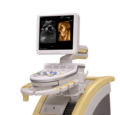
4D Color Doppler Ultrasound
What is the 4D Color Doppler Ultrasound in general?
It is a high-resolution color ultrasound.
The device emits vibrating ultrasound waves towards the body to be imaged and monitors the result for immediate analysis, so that the doctor supervising the procedure sees the desired image immediately so that can make the necessary diagnosis.
Types of cases that are recommended to be diagnosed by 4D Color Doppler Ultrasound:
4D Doppler ultrasound imaging is used to follow up on the condition of fetuses during pregnancy for early diagnosis and is used to depict the state of the live performance of the heart and valves. It is also used to diagnose many possible diseases in other internal organs, to show their organic and practical efficiency within the patient’s body.
How do we do 4D Doppler Ultrasound in a way that distinguishes us from others?
The supervising doctor directs the patient to take the appropriate position to complete the imaging procedure well.
The supervising doctor takes real-time static images to show the necessary scenes to the attending doctor, which is also attached to the final report.
Ecography imaging:
- Imaging of liver, gallbladder, urinary system, and all abdominal viscera.
- Imaging of superficial organs such as (the thyroid – breast – testes & scrotum).
- Doppler imaging of the visceral and superficial vessels (Doppler of the upper and lower extremities, and Doppler of the vessels of the abdomen and liver).
- Color Doppler, flow Doppler of microvessels. It is called “POWER-DOPPLER”.
- Imaging of Visceral elastography, elastography, is a technique that gives a diagnosis of a lesion or mass, whether it is hard or elastic. We can evaluate malignant masses in cases of breast – thyroid – testis – glands.
- Imaging of the salivary glands.
- Echo of the heart: Colored Doppler by a specialist consultant in cardiology.